

Clustered Breast Microcyst
A breast cyst is the most common benign finding in breast cancer screening, and as many as 1/3 of all women will develop a cyst at some point in her life. A cyst is simply an accumulation of fluid within breast tissue, and when it occurs close to the surface and is of enough volume it can form a palpable mass. Sometimes a clinically palpable cyst is called a 'gross cystic mass'. A "Microcyst" on the other hand, is very small, often less than 2-3 mm. They will only show up on a mammogram or ultrasound, and, they often occur in clusters. Breast clustered microcysts are quite common, estimated to appear in around 6% of all breast sonograms.
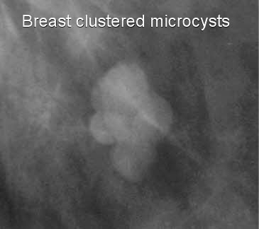
clustered breast microcysts tend to be observed but not treated
Generally speaking, breast clustered microcysts will not be 'treated' per se, as they are vitually always benign, but will be followed up with annual observation.
breast microcysts often show microcalcifications
Microcalcifications are not an uncommon finding with clustered breast microcysts. Certain patterns of microcalcification development have been associated with the early development of ductal carcinoma in situ (DCIS), but the patterns of microcalcification in clustered breast microcysts are very typical, and another indication of a benign situation. Calcifications within clustered breast microcysts tend to develop in 'layers' or 'sedimentation'. When these 'sedimentary' calcifications are discovered on a mammogram, it is even more likely that the radiologist will advise against a biopsy due to the near certainty of a non-malignant cyst.
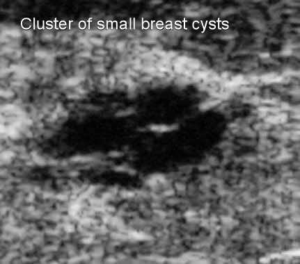
breast lesions with a combination of fluid and solids is a more complex situation
On ultrasound, clustered breast microcysts may appear as tiny anechoic (dark) round areas, (meaning they don't reflect ultrasound waves and don't create echos of sound waves). If the lesion appears to be not entirely made up of fluid, the situation is a bit more complicated. (solid elements might indicate neoplastic cell growth, and would appear to be something more substantial than calcifications.) When the cysts are larger, with 'thicker' portions of the walls, and with indications of some solid particles ( most likely floating debris of various kinds on not malignant cells), the cyst might be described as 'complex', and will likely be biopsied. If some of these more 'complicated' cystic features are present, the radiologist will likely want to make sure that the lesion is not micropapillary ductal carcinoma, which can have a similar appearance. Malignant carcinoma associated with a cyst of microcyst is very unlikely, and usually the comparision of the mammogram and the ultrasound will prove the benign setting. However, any kind of new fibrocystic change in a postmenopausal women (espeically those not taking hormone supplements) will be approached with extra caution, and a peructaneous biopsy is often undertaken.
breast clustered microcysts are probably benign
Clustered breast microcysts would tend to be classified as BI-RADS category 2 to 3 (benign to probably benign), which does indicate a slightly increased risk for potential breast cancer development at some point, but only slightly higher than the general population. When there are no solid components the risk is thought to be very low, and annual follow up is the only necessary course of action.
References
- Berg, WA., Sonographically Depicted Breast Clustered Microcysts: Is Follow-Up Appropriate? AJR 2005; 185:952-959
- Berg, WA., Campassi, CI., Ioffe, OB., Cystic Lesions of the Breast: Sonographic-Pathologic Correlation. Radiology (April 2003) 227, 183-191.
- Demirkazik, FB., Palpable and Nonpalpable Breast Masses. Ultrasound Clinics -(July 2008) Volume 3, Issue 3
- Huff, JG., The Sonographic Findings and Differing Clinical Implications of Simple, Complicated, and Complex Breast Cysts., Journal of the National Comprehensive Cancer Network ,( November 2009) Volume 7 Number 10
- Chang YW, Kwon KH, Goo DE, et al. Sonographic differentiation of benign and malignant cystic lesions of the breast. J Ultrasound Med 2007;26:47–53.
- Stavros AT, Thickman D, Rapp CL, et al. Solid breast nodules: use of sonography to distinguish between benign and malignant lesions. Radiology 1995;196:123–134.
- Kim, KW., Cho, KR, Seo, BK., Whang, KW, Woo, OH., Oh, YW., Kim, YH., Bae, JW., Park, YS., Hwang, CM., Lee, MS., Kim, KI. Sonographic Findings of Mammary Duct Ectasia: Can Malignancy be Differentiated from Benign Disease? J Breast Cancer 2010 March; 13(1): 19-26
- Zhu Q, Cronin EB, Currier AA, et al. Benign versus malignant breast masses: optical differentiation with US-guided optical imaging reconstruction. Radiology. 2005;237:57–66
- Margolin, FR., Jacobs, RP., Denny, SR., Microcystic Calcifications at Stereotactic Breast Biopsy., The Breast Journal (Jan. 2002)
Volume 5 Issue 3, Pages 182 - 185. - Ojeda-Fournier H, Nguyen JQ. How to improve your breast cancer program: Standardized reporting using the new American College of Radiology Breast Imaging-Reporting and Data System. Indian J Radiol Imaging [serial online] 2009 [cited 2010 May 20];19:266-77.
End of page Navigation links: Breast Cancer home or Back to top
Copyright © 1999 - present. Steven B. Halls, MD . 1-780-608-9141 . [email protected]